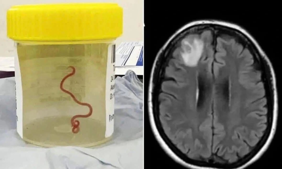
In a first-of-its-kind medical case, doctors in Australia have successfully removed a live, 8-centimetre-long worm from a woman’s brain, a parasite that is normally found in carpet pythons.
The extraordinary case, reported last month by scientists and later shared by Amit Singh Moga, co-founder of The Doctorpreneur Academy, involved a 64-year-old woman who had been suffering from a mysterious illness that baffled doctors for months.

A shocking discovery in the operating room
The woman had undergone a brain scan after experiencing weeks of abdominal pain, diarrhoea, and a persistent dry cough that later progressed to forgetfulness and depression. An MRI showed a lesion in her brain, prompting a biopsy.
“I used tumour-holding forceps and unexpectedly lifted out a thin, squiggling structure,” the neurosurgeon recalled. “My junior doctor initially thought it was an artery, but I pointed out that we were nowhere near one. When I noticed it moving, I immediately asked for it to be removed from my forceps. We placed it into a pathology container and it was still vigorously wriggling.”
The culprit: a python parasite of Australia
Tests later identified the parasite as Ophidascaris robertsi, a roundworm species that usually infects carpet pythons and small mammals. These worms typically complete their life cycle between the two animals, with python faeces containing worm eggs that contaminate grass, which is then eaten by small mammals. The snakes, in turn, consume these mammals, completing the cycle.
In this case, the woman became an accidental host. Living near python habitats, she had foraged native vegetation for cooking, likely ingesting the parasite’s eggs without realising it. Once inside her body, the larvae developed in her intestines before migrating through her bloodstream to her brain.
How the infection affected her
Doctors believe the worm’s movement inside her brain triggered a strong immune reaction, causing inflammation and the formation of a granuloma, a type of lesion often seen when the body tries to isolate a foreign invader. Such brain inflammation can cause a range of neurological symptoms, including headaches, seizures, blurred vision, confusion, or memory loss.
In this case, the woman’s depression and forgetfulness were likely linked to the inflammation caused by the worm’s presence in brain tissue.
Experts say this is the first documented case of a live Ophidascaris robertsi worm being found inside a human brain. Though parasitic infections are not uncommon globally, this discovery has stunned scientists due to the species involved and the unusual route of infection.
The case has also renewed focus on zoonotic diseases, infections that jump from animals to humans and the potential risks of foraging in areas inhabited by wild animals.
While the woman is reportedly recovering, doctors have urged caution in handling or consuming foraged vegetation, especially in areas where wildlife such as pythons are common.
The discovery underscores how close human and animal ecosystems can be and how a simple act like foraging greens from the wild can, in rare cases, lead to medical history being made.
(With inputs from a post by Amit Singh Moga, co-founder, The Doctorpreneur Academy)
Also Read: Did OYO Just Outsmart Its Own Shareholders?